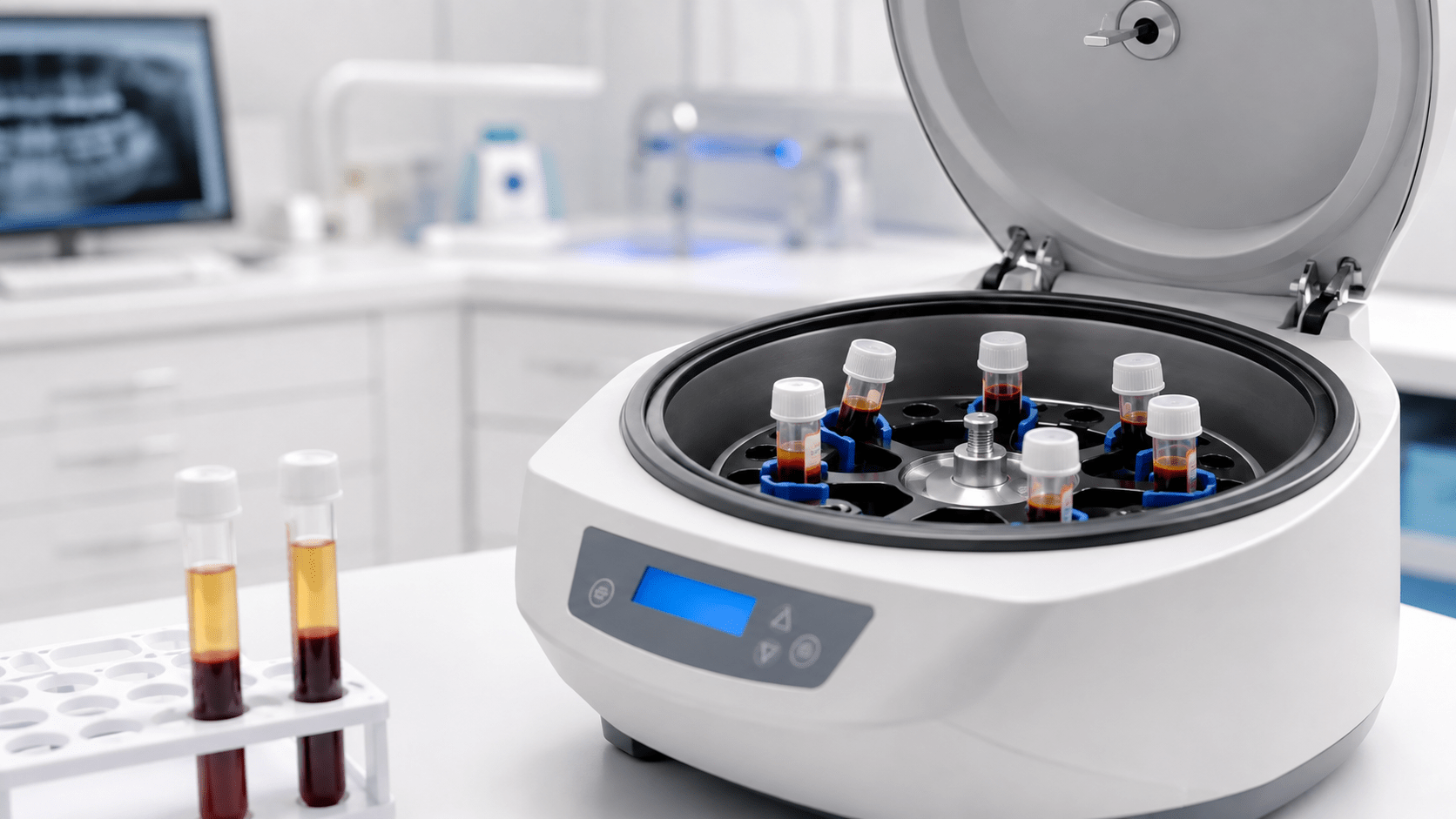
Platelet rich fibrin (PRF) has been utilized in implant dentistry for well over a decade owing to its ability to rapidly stimulate tissue angiogenesis. Over the years, much progress has been made to more effectively concentrate platelets and growth factors as well as define their clinical use for the regeneration of both soft and hard tissues. This product overview will give a brief history of platelet concentrates regarding the advancements made over the years from PRP towards a more autologous three-dimensional scaffold found in PRF. Then, a deep understanding of the years of research leading towards appropriate protocols and methods to best concentrate PRF will be discussed using horizontal centrifugation. Lastly, clinical indications and uses of PRF will be demonstrated with case examples highlighting the use of PRF in extraction site management, bone augmentation, as well as in implant dentistry.
Introduction
Platelet concentrates have been utilized in medicine for over 2 decades owing to their ability to rapidly secrete autologous growth factors and ultimately speed wound healing. They have gained tremendous momentum as a regenerative agent derived from autologous sources capable of stimulating tissue regeneration in a number of medical fields.1-3 Many years ago, it was proposed that by concentrating platelets utilizing a centrifugation device, growth factors derived from blood could be collected from a platelet-rich plasma layer, and later utilized in surgical sites to promote local wound healing.2,3 Today, it has been well established that platelet concentrates act as a potent mitogen capable of:
- Speeding the revascularization of tissues (angiogenesis)
- Acting as a potent recruitment agent of various cells including stem cells
- Inducing the prompt multiplication of various cell types found in the human body (proliferation)
Specifically in dentistry, platelet concentrates where introduced over 20 years ago by Marx and colleagues with the aim of concentrating blood proteins as a natural source of growth factors that could stimulate vascularization (angiogenesis) and tissue ingrowth based on the fact that blood supply is pivotal for tissue regeneration of all tissues.4 Wound healing has been described as a 4 step process which includes 1) hemostasis, 2) inflammation, 3) proliferation, and 4) maturation.5-7
The Evolution of Platelet Rich Fibrin
It is interesting to point out that the use of platelet concentrates has slowly and gradually gained in popularity with a dramatic increase being observed in the past 5-10 years. Platelet-rich plasma (PRP), as its name implies, was designed to accumulate platelets in supra-physiological doses within the plasma layer following centrifugation. The main aim of PRP was to isolate and further concentrate the highest quantify of platelets and their associated growth factors for regenerative purposes and thereafter re-implanting this specialized supra-concentrate at sites of local injury.1
Initial protocols typically ranged in duration from 30 minutes to 1 hour based on the centrifugation/collection systems and protocols utilized. Since lengthy protocols were utilized, anti-coagulants were added to blood collection tubes. These typically were either various forms/concentrated bovine thrombin and calcium chloride.
Despite its growing success and continued use, several reported limitations existed with respect to reaching its full healing potential. Mainly, the use of anti-coagulants was shown to limit wound healing.1 Simply put, when injury occurs leading to an open wound, a blood clot is one of the first and most crucial steps that occurs in order for healing to take place. Shortly thereafter, cells and growth factors get trapped within this newly formed extracellular matrix and the wound healing process/cascade begins. By limiting the body’s ability to form a clot-stable tissue, wound healing is delayed. Several studies have now demonstrated the superior outcomes of PRF when compared to PRP simply by removing anticoagulants from their formulations.1
Another drawback of PRP was the fact it remained liquid by nature (use of anti-coagulants) and therefore when it was typically combined with biomaterials. When compared to PRF, PRP demonstrates a quick initial burst of growth factors being released whereas PRF demonstrates a more slow and gradual release of growth factors over an extended period of time that has since been shown to significantly improve cell growth and tissue regeneration.8,9
Leukocyte and Platelet Rich Fibrin
Owing to the main drawback that anticoagulants utilized in PRP prevented clotting, platelet rich fibrin (PRF) was developed with the main aim of simply removing anti-coagulants.10 By doing so, a much quicker working time was needed and the practitioner absolutely required centrifugation to begin shortly after blood collection (otherwise the blood would naturally clot within a tube). The main advantage of this fibrin matrix is the ability for it to release growth factors over an extended period of time while the fibrin clot is being degraded (as opposed to PRP which remained in liquid form having a much faster growth factor release profile).11 Over the years, PRF has also been termed L-PRF (for leukocyte and platelet rich fibrin) owing to the discovery that several leukocytes remained incorporated in PRF.
Recently, a series of basic laboratory experiments revealed better means to optimize the production of PRF using horizontal centrifugation. Simply, horizontal centrifuges are routinely utilized in high-end research labs as well as in medical hospitals owing to their greater ability to separate layers based on density. Unlike fixed-angle centrifugation system whereby the tubes are actually inserted on a ~45 degree angle, horizontal centrifugation (often referred to as swing-out bucket centrifugation) have the ability for tubes to swing-out to 90 degrees once they are in rotation. This technology leads up to 4 times greater cell content when compared to fixed-angle centrifugation.12 The major disadvantages of fixed-angle centrifugation is that during the spin cycles, cells are typically driven along the back wall of centrifugation tubes at high g-forces with relative difficult to separate properly according to their cell density. This also exposes cells to higher compressive forces against the back wall and cells must then separate by travelling either up or down the inclined centrifugation slope based on their respective cell density differences. Since red blood cells are larger and heavier than platelets and leukocytes, they travel downwards, whereas lighter platelets travel towards the top of the tube where PRF is collected. This makes it relatively difficult for the smaller cell types such as platelets and particularly leukocytes to reach the upper, especially granted that RBCs outnumber in particular WBCs typically by ~1000 fold. Therefore and in summary, by utilizing a fixed-angle centrifuge, it is not possible to reach optimal accumulation of platelets or leukocytes as a result of their fixed-angle system design.
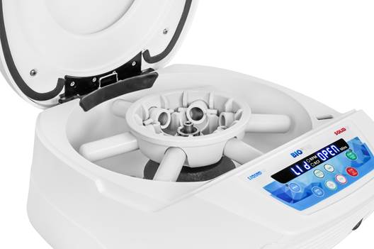
Figure 1: Clinical photo of a BIO-PRF centrifuge. Photo demonstrating the horizontal centrifugation concept. When tubes are inserted vertically (up and down), once the device begins to rotate the tubes swing out completely horizontally. This favors better blood cell layer separation with higher platelet and growth factor concentrations. Reprinted with permission from Miron 2021.
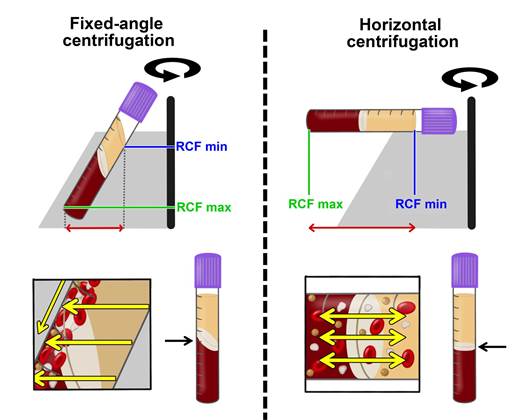
Figure 2: Illustrations comparing fixed-angle and horizontal centrifuges. With horizontal centrifugation, a greater separation of blood layers based on density is achieved owing to the greater difference in RCF-min and RCF-max. Following centrifugation on fixed-angle centrifuges, blood layers do not separate evenly, and as a result, an angled blood separation is observed. In contrast, horizontal centrifugation produces even separation. Owing to the large RCF values (~200-700 g), the cells are pushed toward the outside and downwards. On a fixed-angle centrifuge, cells are pushed toward the back of centrifugation tubes and then downwards/upwards based on cell density. These g-forces produce additional shear stress on cells as they separate based on density along the back walls of centrifugation tubes. In contrast, horizontal centrifugation allows for the free movement of cells to separate into their appropriate layers based on density, allowing for better cell separation as well as less trauma/shear stress on cells. Modified from Miron et al. 2019.
Dental Uses of PRF
In dentistry, the majority of procedures are done utilizing the 700 RCF for 8 minute protocol. Specifically to GBR, the so-called ‘sticky bone’ protocol utilized this spin rate when both liquid-PRF (blue or white) and solid-PRF (red) tubes are drawn simultaneously and centrifuges at the same time. Always remember to draw liquid-PRF tubes first. While an entire textbook has been dedicated on this topic, presented are a few cases of use of PRF for 1) extraction site management, gingival recession coverage, and in implant dentistry to support soft tissue healing and reduce bacterial infiltration.
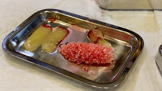
Figure 3: Clinical photo of a fabricated ‘sticky bone’ by using particulate allograft mixed with Solid-PRF fragments (cut up PRF membranes) with the addition of liquid-PRF. Reprinted with permission from Miron 2021.
