Platelet-rich fibrin (PRF) has been demonstrated to be an excellent biomaterial addition to most surgeries performed in dentistry owing to its excellent biocompatibility, super-concentrate of autogenous cells, and release of regenerative growth factors over time. Nevertheless one of the main reported drawbacks of PRF is its rather fast resorption properties characterized within a 2-3 week timeframe, thus it could never be utilized as a ‘barrier’ membrane in GBR/GTR procedures. Interestingly, recent studies have demonstrated that by heating a liquid platelet-poor plasma (PPP) layer, the resorption properties of heated albumin (albumin gel) can be significantly extended from 2 weeks to greater than 4 months (extended-PRF; e-PRF) using the Bio-Heat technology. This product report highlights the scientific development from 2019 until today whereby this novel regenerative modality demonstrates for the first time the marked improvement in membrane stability of these extended/heated-PRF membranes. These e-PRF membrane have since been utilized as a biological barrier membrane for GBR procedures with a longer-lasting half-life, or as a biological filler material in esthetic medicine applications.
Introduction
Platelet concentrates have been utilized in many fields of regenerative medicine owing to their ability to deliver supraphysiological concentrations of autologous platelets, leukocytes and growth factors.1,2 While platelet-rich plasma (PRP) has been extensively employed as a first generation platelet concentrate, platelet rich-fibrin (PRF) has since been utilized with a better ability to release more growth factors over an extended period of time with superior clinical outcomes in various fields of medicine and dentistry.3,4 PRF has proven to be an excellent biomaterial composed of autogenous cells and growth factors entrapped within a fibrin network that have been shown to breakdown more slowly over time when compared to traditional PRP.4
Nevertheless, one of the main reported drawbacks of PRF (and especially PRP) is its faster-than-ideal resorption rate characterized within a 2-3 week timeframe.5 Because of its fast resorption rate, PRF membranes cannot be utilized as a barrier membrane similar to collagen owing to their inability to exclude soft tissues over an extended period of time. Interestingly, a number of recent studies have demonstrated that PRF could be significantly extended from 2-3 week resorption properties to greater than 4 months (extended-PRF; e-PRF) by heating a liquid platelet-poor plasma (PPP) layer (denaturing albumin) using Bio-Heat technology.6
The heated version of plasma, which has now been utilized in many areas of medicine and dentistry, has recently been the basis of intensive research endeavors across many regenerative labs owing to its extended working properties. Since one of the main limitations of PRP/PRF has historically been its short in vivo turnover rate, these extended PRF membranes can be used as substitutes for collagen membranes in guided bone regeneration (GBR) procedures requiring a typical ‘barrier’ function that protects bone regeneration from faster growing soft tissues.7
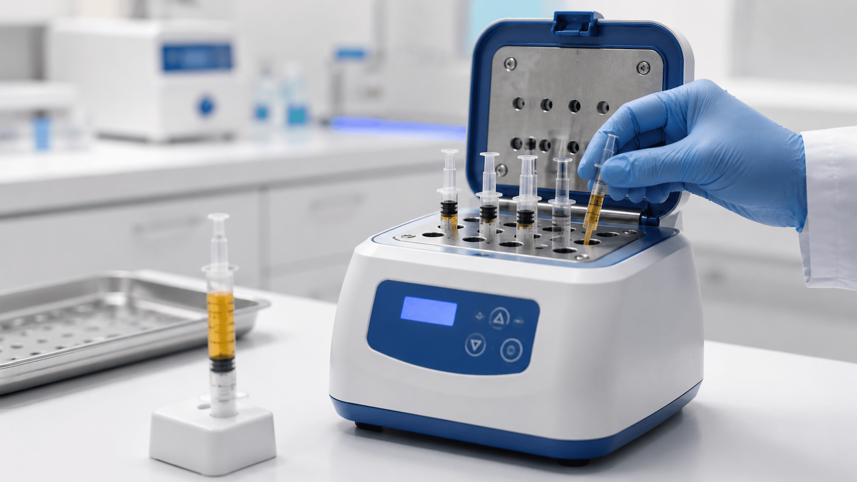
To overcome the quick degradation properties of platelet concentrates and better maintain volume stability, the Alb-PRF protocol was developed. This protocol involves the use of the PPP layer (containing ~60% albumin) and heating it to 75 °C for 10 minutes to allow for albumin denaturation as well as breaking of many weak linkages or bonds (e.g., hydrogen bonds) within its protein molecule. Following denaturation, the proteins are then restructured in a more densely organized assembly that extends the resorption properties of PRF to up to 4-6 months.8-11 However, the heating process also destroys cells/growth factors, and thereby, the platelet concentrates lose much of their regeneration potential. For these reasons and following heating, a concentrated PRF layer (C-PRF)12,13 taken from the buffy coat is then mixed back into the heated PPP (albumin gel) once cooling has occurred (termed Alb-PRF or e-PRF for extended PRF).14
In view of previous research to date on this topic, the aim of this product review was to present the biological properties of this novel regenerative modality. Furthermore, the step-by-step processing of e-PRF as well as clinical studies to date on the topic is presented in Figure 1. As an injectable filler, a 25G needle or 22G canula is recommended. Alternatively, a similar process can be utilized to create a custom shape membrane with extended properties (e-PRF) lasting up to 4 months and utilized clinically as a substitute for collagen membranes (Figure 2).
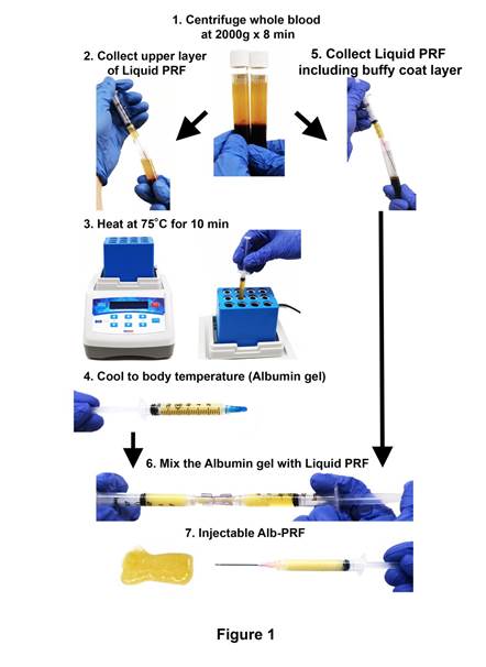
Figure 1: e-PRF preparation protocol. 1) Whole blood was centrifuged at 2000 × g for 8 minutes. The upper layer (yellow layer) shows the liquid plasma layer. 2) The uppermost layer of platelet-poor plasma (PPP) was collected in a syringe. 3) The collected PPP was heated in a heat block device at 75°C for 10 minutes and thereafter 4) cooled to room temperature for approximately 10 minutes. An injectable albumin gel was then prepared. 5) The liquid platelet-rich layer (liquid-PRF), including the buffy coat layer with accumulated platelets and leukocytes, was collected in a separate syringe. 6) The albumin gel and native liquid PRF were then thoroughly mixed by utilizing a female-female luer lock connector. 7) Injectable e-PRF in final ready form. Reprinted with permission from Fujioka-Kobayashi et al. (2020).
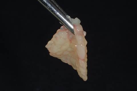
Figure 2: An e-PRF membrane prepared for use. Case performed by Dr Ezio Gheno. Reprinted with permission from Miron 2021.
Clinical Use of Alb-PRF/e-PRF Membranes for Periodontology and Implant Dentistry
One of the main areas where the e-PRF membranes were first clinically utilized and tested was as a substitute for collagen membranes. Therefore, several clinical case series were started whereby the e-PRF membranes were created and utilized for extraction site management to cover bone allografts following tooth removal similar to either a collagen membrane/plug or a PTFE membrane (Figure 3). This opportunity offers the clinician a 100% all-natural biomaterial with extended resorption properties that can also be left exposed in the oral cavity following placement. Notably, the e-PRF membranes may also be left exposed in the oral cavity, and their inclusion of supra-physiological concentrations of leukocytes allows for greater defense against incoming pathogens. Figure 3F highlights the much faster soft tissue closure ability of epithelial tissue over the e-PRF membrane when compared to alternative strategies.
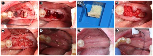
Figure 3: Clinical case using an e-PRF membrane as a substitute for a collagen barrier membrane following extraction site management. (a) Sectioning of teeth. (b) Following tooth extraction. (c) Allograft bone grafting with PRF fragments. (d) Fabrication of the e-PRF membrane following heat treatment at 75 °C for 10 minutes in the Bio-Heat device. (e) e-PRF membrane in place with suturing. (f) One week postoperatively. Note the excellent healing. (g) 8 weeks post-op. (h) 4 months post-op. Case performed by Dr Nathan Estrin.
E-PRF membranes have been utilized extensively during GBR procedures (Figure 4). Here, such membranes may be much less foreign to soft tissues, with reports of improved biocompatibility. Finally, e-PRF membranes have also been utilized as a substitute for various collagen membranes during recession coverage procedures utilizing a vestibular tunneling approach (Figure 5).
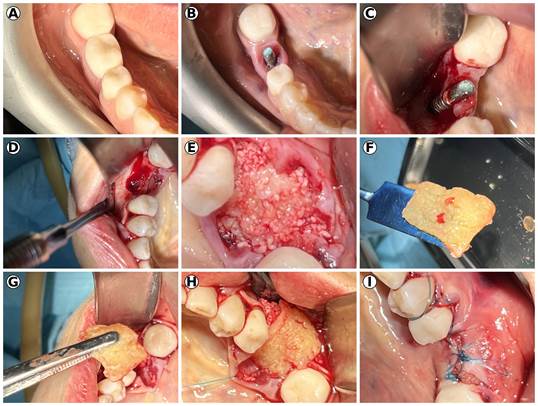
Figure 4: Use of the e-PRF membrane for GBR. (A, B) Lower first molar demonstrating deficiency of the buccal surface of the implant. (C) Flap elevation demonstrating complete loss of the buccal bone of this malpositioned implant. (D) Implant removal. (E) Grafting of the defect site with bone particles mixed with PRF fragments. (F) Fabrication of an e-PRF membrane. (G, H) Placement of the e-PRF membrane covering the grafted defect. (I) Final soft tissue closure. Case performed by Dr. Alan Rene Espinoza.
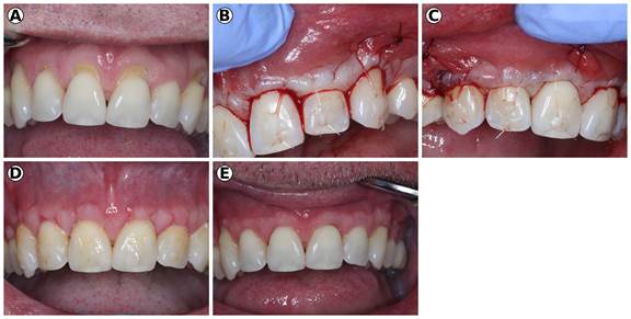
Figure 5: Use of e-PRF for the management of multiple gingival recessions. (A) Preoperative images demonstrating multiple anterior gingival recessions. (B-C) Postsurgical result following tunneling with e-PRF via a vestibular incision. (D) Two weeks post-op; note the excellent soft tissue healing. (E) 8 weeks post-op. Case performed by Dr Nathan Estrin.
Clinical Studies Using e-PRF as an Injectable Bio-Filler
To date, thousands of cases have utilized the technology of extending the working properties of platelet concentrates as a biological filler (Bio-Filler). One of the first approaches was to restore the facial volume (Figure 6), Interestingly, Alb-PRF has been utilized for injections into deficient papilla with quite promising results, yielding up to 4 years of data (Figure 7). Last, Alb-PRF has also been utilized as a biological growth factor for joint and spinal injections such as diseased osteoarthritic joints and spinal injections.
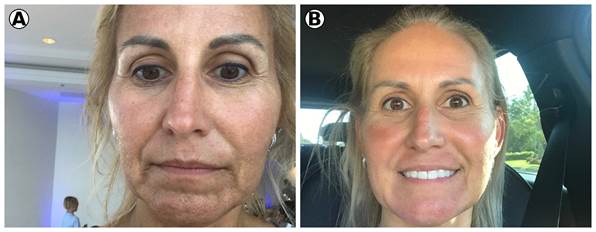
Figure 6: (A) Mid-40-year-old female patient with pronounced marionette lines, deep nasolabial folds and an overall aged facial appearance. (B) Final outcome following 3 treatments with 100% natural approaches, including laser therapy, microneedling with PRF, and e-PRF injections. Case performed by Dr. Scott Delboccio. Reprinted with permission from Miron 2021.
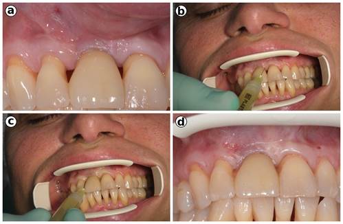
Figure 7: a) Clinical photograph demonstrating maxillary anterior teeth with multiple black triangles. (b-c) Injection techniques of the Alb-PRF mixture into various papillas. (d) Final clinical photographs after injections. Case performed by Dr Ezio Gheno. Reprinted with permission from Miron 2021.
