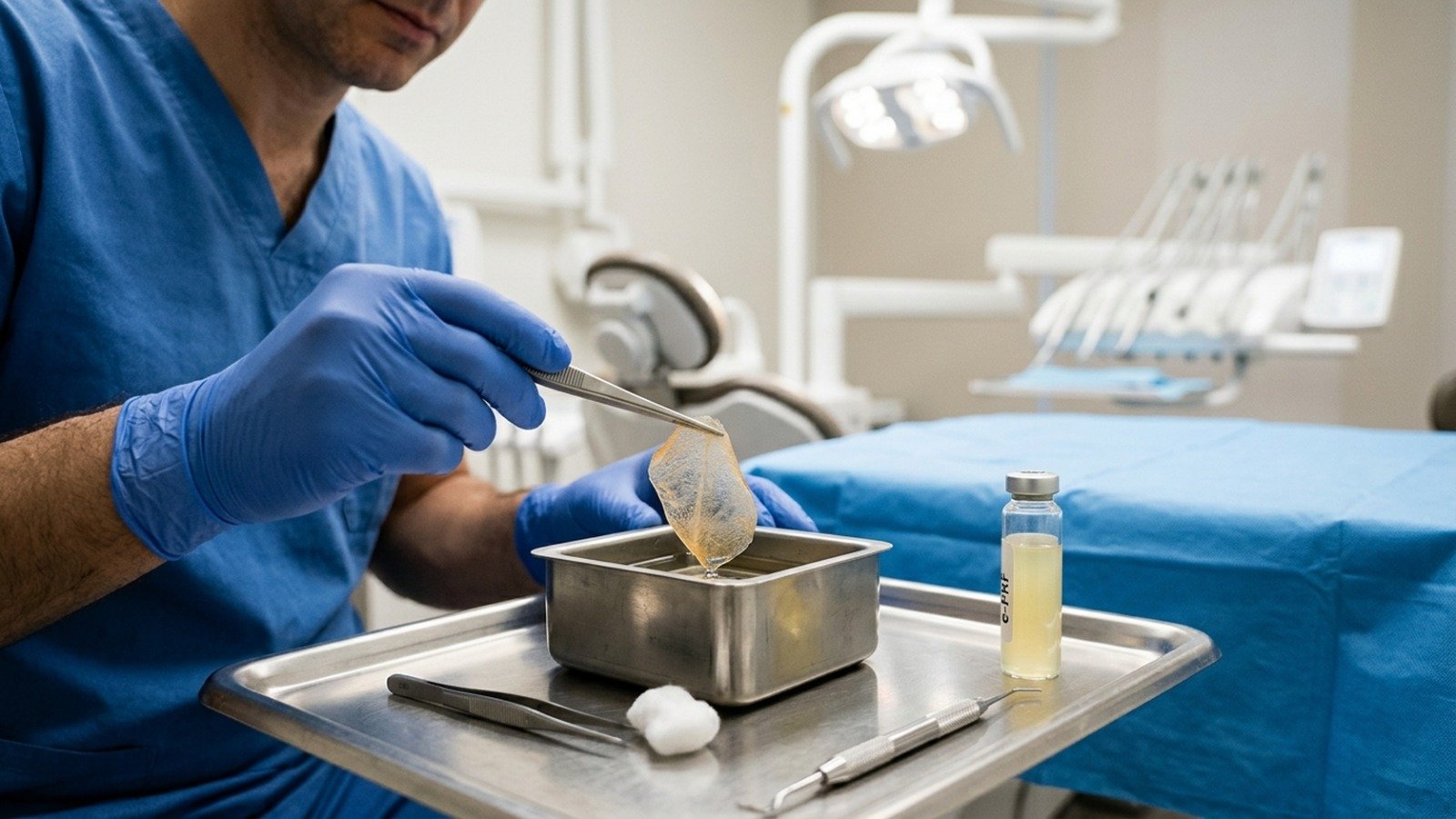
Platelet-rich Fibrin (PRF) is an autologous regenerative biomaterial that is utilized in dentistry for well over a decade. PRF has emerged as a superior platelet concentrate over platelet-rich plasma (PRP) due to its increased concentration of growth factors by eliminating anticoagulants in the collection tubes. Utilizing hydrophilic glass tubes, and hydrophic plastic tubes, PRF can be created and utilized in both solid and liquid form respectively. In the case of solid-PRF, a fibrin clot is created which releases growth factors continuously over time as it resorbs. One disadvantage however that prevents utilizing this solid-PRF as a membrane in ridge preservation procedures is the fast resorption time which has been characterized in the literature as lasting only 2-3 weeks. If the membrane has resorbed prior to complete wound closure, the grafted socket is left vulnerable. A novel therapy introduced only recently has demonstrated that by heating PRF via utilization of the BioHeat technology, PRF and more specifically albumin proteins found in plasma can be denatured which dramatically extends the resorption properties of PRF by 4-6 months. This novel formulation of PRF has since been termed ‘extended’-PRF or simply ePRF. In the clinical case presented herein, this innovative ePRF has been utilized as a solo membrane in ridge preservation of tooth #20 successfully leading to implant placement in a healthy 67-year-old female. This novel technology presents many new opportunities for implant surgeons by providing them with the ability to create all-natural, low-cost, and extremely biocompatible barrier membranes without the use of a single chemical or biomaterial derived 100% from the patient’s own blood.
Introduction
Platelet-rich Fibrin (PRF) is an autologous regenerative biomaterial that is being increasingly utilized due to continuous improvements in centrifugation methods, tube quality, and clinical applications.1 PRF has emerged as a superior platelet concentrate over the first generation platelet-rich plasma (PRP) due to its increased release of growth factors over time by eliminating anticoagulants in the collection tubes.2,3 A blood clot is one of the first and most important steps in wound healing and introducing anti-coagulants impedes the process of forming a complete fibrin extracellular matrix thus limiting wound healing potential.4,5 Several studies have demonstrated the increased concentration of growth factors in PRF when compared to PRP and even the original research group that developed the idea of plasma rich in growth factors (PRGF) has since demonstrated superior healing potential with anti-coagulant removal.6 PRF includes a shorter preparation time and clinical protocols are faster and easier for the clinician to implement in order by successfully removing anti-coagulants. Due to the shorter working time during blood harvesting, clinicians will need to centrifuge the blood shortly after collection to avoid natural blood clotting within the tube. Utilizing hydrophilic glass tubes, and hydrophic plastic tubes, PRF can be created and utilized in both solid and liquid form respectively with the exact same centrugation protocol.1 While the liquid form of PRF allows for an injectable mode of delivery, solid-PRF is a fibrin matrix with an ability to release growth factors continuously as the clot degrades.5 One disadvantage from utilizing this solid-PRF as a membrane in ridge preservation procedures however is its fast resorption time of 2-3 weeks.7 If the membrane has resorbed prior to complete wound closure, the grafted socket is left vulnerable. Therefore, it is not an ideal choice of biomaterial for ridge preservation prior to implant therapy.
Due to the high success rate and ideal esthetic and functional outcomes, dental implants remain the gold standard for the replacement of missing teeth.8 Successful implant placement begins with managing the hard and soft tissue dimensional changes that occur after dental extractions. Ridge preservation has been achieved using a combination of bone grafts, membranes, and growth factors to minimize post-extraction dimensional changes.9 PRF has established itself as an essential component of the processes involved in ridge preservation as it favors faster revascularization of defect areas and provides growth factors capable of recruiting progenitor cells for healing. Most commonly, both solid and liquid PRF are mixed with bone particulate to create “sticky bone” for easier handling properties and adding growth factors to the boney scaffold.10 Additionally, solid-PRF is often utilized as a membrane layered over a traditional collagen resorbable membrane. But as previously mentioned, its fast resorption time discourages it’s use as a sole membrane. Interestingly, the Bio-Heat technology developed recently, has demonstrated that by heating PRF, the resorption properties of PRF can be dramatically extended from 2-3 weeks to 4-6 months (Figure 1).11 Therefore, this novel extended platelet-rich fibrin (ePRF) has potential to be utilized as a membrane alone, completely replacing the need and use of collagen membrane (Figure 2).12 In the clinical case presented below, an ePRF membrane was utilized as an unaccompanied membrane in the ridge preservation of tooth #20 successfully leading to implant placement in a healthy 67 year old female.
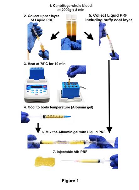
Figure 1: e-PRF preparation protocol. 1) Whole blood was centrifuged at 2000 × g for 8 minutes. The upper layer (yellow layer) shows the liquid plasma layer. 2) The uppermost layer of platelet-poor plasma (PPP) was collected in a syringe. 3) The collected PPP was heated in a heat block device at 75°C for 10 minutes and thereafter 4) cooled to room temperature for approximately 10 minutes. An injectable albumin gel was then prepared. 5) The liquid platelet-rich layer (liquid-PRF), including the buffy coat layer with accumulated platelets and leukocytes, was collected in a separate syringe. 6) The albumin gel and native liquid PRF were then thoroughly mixed by utilizing a female-female luer lock connector. 7) Injectable e-PRF in final ready form. Reprinted with permission from Fujioka-Kobayashi et al. (2020).
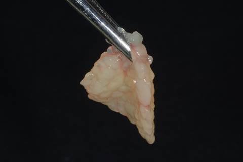
Figure 2: An e-PRF membrane prepared for use. Case performed by Dr Ezio Gheno. Reprinted with permission from Miron 2021.
Case Description
A 67-year-old female reported with a hopeless tooth #20 due to a vertical root fracture. Outside of well-managed hypertension, the patient’s medical history was insignificant. The patient was treatment planned for delayed implant placement 3 months following ridge preservation. Figure 3A shows a preoperative clinical photograph. After atraumatic extraction of tooth #20, the socket was degranulated with a spoon excavator leaving a 4-wall socket and a thin buccal plate (Figure 3B). By mixing both solid and liquid-PRF with particulate bone allograft, “sticky bone” was created and packed in the socket (Figure 3C). To fabricate the e-PRF membrane, a standard protocol was followed according to those previously published.13 Briefly, a 21G butterfly needle was utilized to draw two 10ml liquid-PRF tubes of blood from the patient and centrifuged at 700g for 8 minutes utilizing a horizontal centrifuge (Bio-PRF, Florida, USA). After centrifugation, one syringe was utilized to draw 2-4mL of the platelet poor plasma (PPP) layer from one blue tube and placed in the Bio-Heat Device for 10 minutes at a temperature of 75 degrees Celsius for serum albumin denaturation. Following this protocol, liquid-PRF converts into a plasma gel with a 4-6 month resorption time. A second syringe was then utilized to draw 2-4mL of liquid PRF from the buffy coat zone. The denatured albumin gel was then placed and condensed in a custom Bio-Heat tray to the desired shape. The liquid PRF from the buffy coat was then removed and utilized to saturate the albumen gel in the Bio-Heat tray re-introducing growth factors and clotting factors within the final ePRF membrane composition. The gel and liquid-PRF were then left for 10 – 15 minutes to set prior to use. Once completed, the ePRF membrane was placed over the grafted socket (Figure 3D) and sutured with 4-0 chromic gut in mattress fashion (Figure 3E). The patient was brought back for a 2-week post-operative evaluation displaying complete wound closure (Figure 3F). At the 3-month evaluation, a CBCT was taken displaying adequate bone for implant placement (Figure 3G). Upon implant placement, notice the contour of the ridge was well maintained with excellent bone quality (Figure 3H). a 3.4 x 10.5mm BioHorizons Tapered Pro implant was placed (Figure 3I). Due to sufficient torque value, a healing abutment was placed, and final suturing completed with 4-0 chromic gut in single interrupted fashion (Figure 3J).
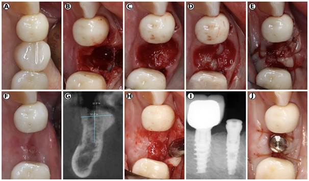
Figure 3: A) Preoperative photograph of #20 prior to extraction. B) Extraction socket with thin buccal plate. C) “Sticky bone” packed in the socket created with particulate bone allograft and both solid and liquid-PRF. D) An extended-PRF (ePRF) membrane created utilizing liquid PRF and the BioHeat Technology to secure the membrane. E) Final suturing with 4-0 chromic gut in mattress fashion. F) 2-week post-operative healing displaying complete wound closure. G) CBCT cross-sectional view showing adequate bone for implant placement. H) Flap reflection at 3-months displaying adequate bone regeneration for implant placement. I) Periapical radiograph of Implant placement (3.4 x 10.5mm BioHorizons Tapered Pro Implant) J) Implant placement with Healing Abutment and 4-0 chromic sutures in single interrupted fashion. Case performed by Dr. Nathan Estrin.
Conclusion
This case report demonstrated a successful outcome utilizing the novel extended platelet rich fibrin as solo membrane in ridge preservation. The ability to utilize the patient’s autologous blood as a replacement to collagen membrane may provide both an economical and biological advantage over traditional collagen membranes. While more case series and cross-sectional studies are required to further validate its use, the utilization of Bioheat technology to construct the unique ePRF membrane appears to be a viable tool in bone grafting techniques and implant dentistry in the future. Furthermore, additional studies utilizing the e-PRF in commonly utilized collagen membrane applications, such as GBR, sinus grafting, and recession coverage, also merits further investigation.
